Glutathione detection and imaging technology
Li Lingling's research team at Nanjing Medical University published an important research result in the international journal Sensors and Actuators: B. Chemical (Sensors Actuators B Chem. 2024, 403, 135200).
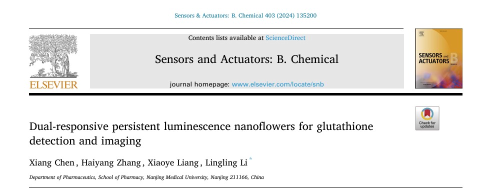
The research team designed and synthesized a dual-mode sensor (ZCGO@MnO₂) based on sustainably glowing nanoflower flowers that can be used for high-sensitivity detection and imaging glutathione (GSH).
This achievement provides a new technical solution for the field of biosensing, and shows a broad application prospect in disease diagnosis and cell status monitoring.
Background introduction
Glutathione (GSH) is a key molecule in maintaining REDOX balance in vivo and is involved in cell signaling and protein function regulation.
Abnormal levels of glutathione are closely associated with a variety of diseases, such as neurodegenerative diseases, cancer, cardiovascular disease, etc.
Drug-induced changes in glutathione may also lead to cytotoxicity and apoptosis, so real-time monitoring of glutathione levels is of great significance for disease diagnosis and evaluation of drug efficacy.
Current techniques for glutathione detection include fluorescence, electrochemical and colorimetry.
These methods are often affected by background spontaneous fluorescence interference, resulting in a low signal-to-noise ratio (S/N), especially when applied in complex biological substrates.
Continuous luminescent nanoparticles (PLNPs) have become a research hotspot in the field of biosensing because they can emit light without real time excitation and can effectively eliminate background interference.
Traditional PLNPs has uneven structure and difficult surface modification, which limits its application in multi-mode sensing.
Main research content
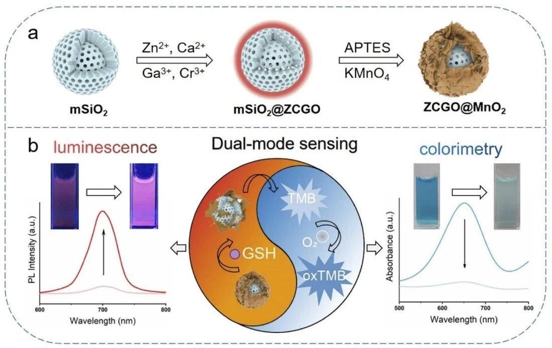
In this study, we developed a sensor based on PLNPs and MnO₂ bi-functional nanostructures, which combined continuous luminescence and colorimetric signals to achieve two-mode glutathione detection.
Through template-assisted synthesis, the research team prepared homogeneous MSIO2 @ZCGO nanoparticles, and further modified by APTES and deposited MnO₂ layer to form a unique nanoflower structure.
As an energy receptor, MnO₂ quenches the luminescence of PLNPs through the luminescence resonance energy transfer (LRET) mechanism, and exhibits oxidase-like activity, which can catalyze TMB to produce blue oxidation products.
In the presence of glutathione, the MnO₂ layer is reduced to Mn² +, resulting in the restoration of luminescence and inhibition of enzyme-like activity.
The experimental results show that the sensor has excellent linear detection performance for glutathione in the range of 5-200 μM, and the detection limit is as low as 0.33 μM.
The signal variation in PL mode eliminates the complex matrix background interference and significantly improves the detection sensitivity and reliability.
The selectivity and stability of the sensor are also verified. The sensor is insensitive to interference from other amino acids and metal ions, and only responds significantly to biothiols such as Cys and glutathione.
The sensor maintained stable performance for 14 days, proving its viability for long-term applications.

(a) UV-VIS absorption spectra of the reaction system at different concentrations of glutathione;
(b) Linear fitting curve between absorbance change and glutathione concentration;
(c) Selective experimental results of colorimetric detection
Through cell imaging experiments, the team further demonstrated that the sensor could be used to monitor glutathione levels in living cells.
Imaging results of cancer cells and normal cells showed that the concentration of glutathione in cancer cells was significantly higher than that in normal cells, consistent with theoretical expectations.
Through drug treatment experiments, the study revealed the change law of glutathione level with the action of drugs, indicating that the sensor has the power to evaluate the efficacy of drugs.
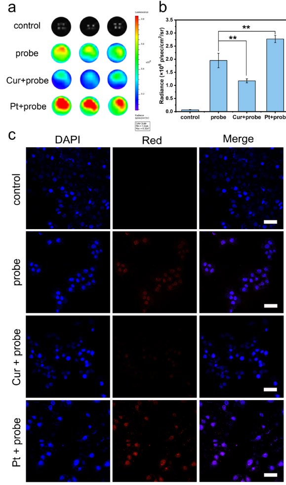
(a) Continuous luminescence imaging of MCF-7 cells after 2 hours without or with the probe after different pretreatments;
(b) Quantitative analysis of PL signals under different preprocessing conditions, with data expressed as mean ± standard deviation;
(c) confocal microscopic imaging results of ZCGO@MnO₂ in MCF-7 cells under different preconditioning conditions.
conclusion
The dual-mode continuous luminescence sensor developed in this study realizes efficient detection and imaging of glutathione by combining LRET and enzyme-like catalytic mechanism.
This research achievement not only provides a reliable means for the accurate detection of glutathione in complex biological environments, but also broadens the application range of PLNPs in the field of biosensing and imaging, and provides a strong technical support for future disease diagnosis and drug research.
The research team plans to further optimize the sensor's performance and explore its potential applications in other biomarker detection.
Data source: https://doi.org/10.1016/j.snb.2023.135200


